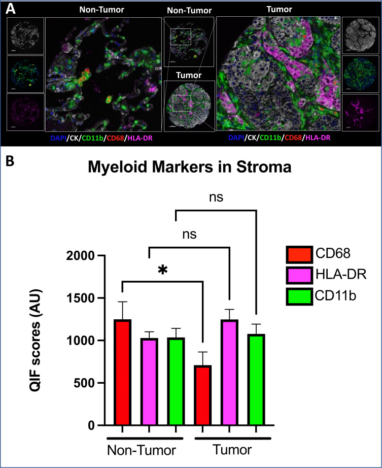
Figure 1.
Decreased levels of differentiated myeloid cells in tumor versus non-tumor samples. (A) Representative images of non-tumor (left) and tumor (right) with respect to CD11b, CD68, and HLA-DR expression. (B) Statistically significantly higher QIF level of differentiated myeloid cell marker (CD68) in non-tumor than in tumor cells (p=0.0165). AU, AQUA units; CK, cytokeratin; DAPI, 4’,6-Diamidino-2-Phenylindole; QIF, quantitative immunofluorescence.

