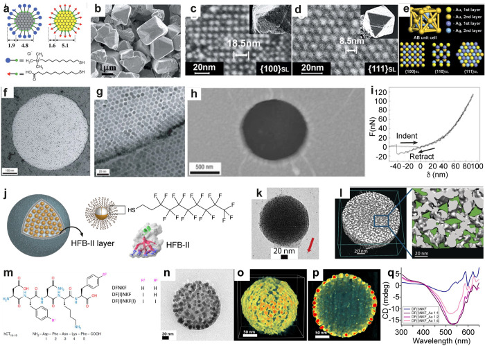
Figure 3.
Self-assembled 3D crystals, 2D arrays, and spherical particles using narrow size dispersed nanoparticles. (a–e) Schematics of AuMUA and AgTMA and SEM, HRSEM, and scheme of diamond-like crystals with the projections of {100}SL, {110}SL, and {111}SL planes, respectively. (f–i) TEM images, AFM image, and force vs. distance curve showing elasticity of the self-assembled dodecanethiol capped AuNP membrane. (j–l) Schematics, TEM image (red arrow indicates fiducial gold markers), and 3D-reconstructed electron tomogram, respectively, of fluorinated supraparticles assembled using HFB-II. (m) Chemical structure of iodinated peptides used for in situ NP formation. (n–p) TEM image, 3D-reconstructed tomogram, and cross-sectional view showing monolayer shell of AuNPs and unreacted peptides in the core, respectively. (q) CD spectra of DF(I)NKF with varying ratio of Au. Panels a–e reproduced with permission from ref (7). Copyright 2006 AAAS. Panels f–i reproduced with permission from ref (9). Copyright 2007 Springer Nature Ltd. Panels j–l reproduced with permission from ref (17). Copyright 2017 John Wiley & Sons. Panels m–q reproduced with permission from ref (18). Published 2019 by American Chemical Society.

