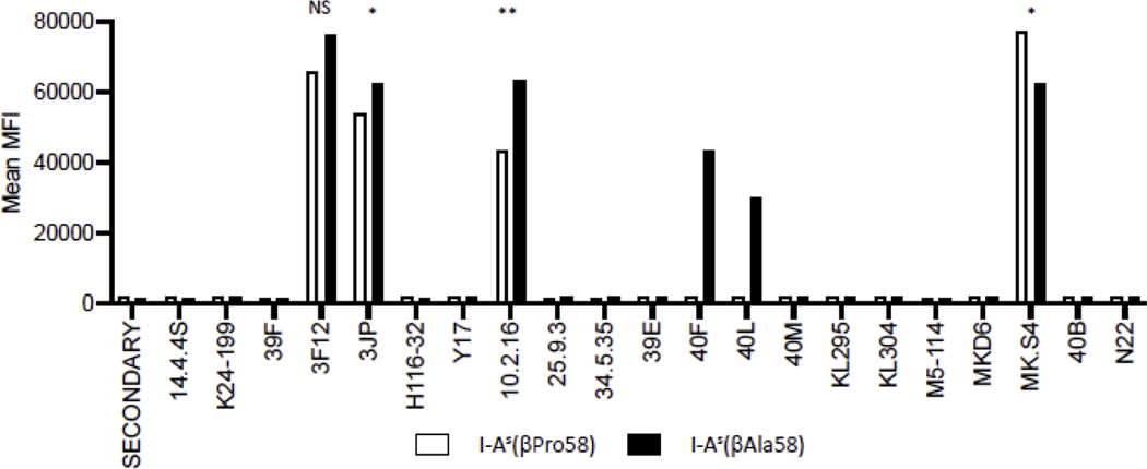
Figure 2. Reactivity of APC expressing either I-As(βPro58) or its variant I-As(βAla58) with monoclonal antibodies (mAb).
Transfectants expressing either I-As(βPro58), white bars, or I-As(βAla58), black bars, were stained with the indicated mAb and then mean fluorescence intensity (MFI) of the population, which was >90% positive, was scored by flow cytometry in a two-step staining procedure. The MFI is indicated for each monoclonal antibody. Shown here is a representative figure. Each antibody was tested in at least two independent experiments. Statistical analysis was completed for mAb with reactivity to both MHC class II molecules, using an unpaired t-test.

