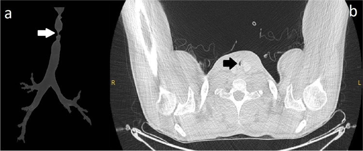
Fig. 1.
Computed tomographic view of the tracheal stenosis. a 3-Dimensional reformation of the trachea. The narrowest tracheal segment was marked with a white arrow. b Axial plain computed tomography reveals the narrowest tracheal segment (marked with black arrow). The patient was relieved symptomatically with tracheal dilatation, and then, elective tracheal resection and reconstruction was performed

