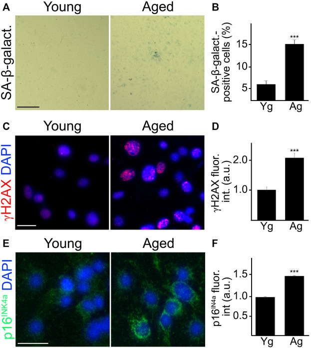
FIGURE 1.
Cultured primary CEC from aged mice recapitulated senescence-associated phenotypes observed in vivo. CEC were isolated from young (3–4 m/o) and aged (18–20 m/o) female mice and cultured. Young and aged cultured CEC were stained for SA-β-galactosidase (A), with antibodies against the double-strand DNA damage marker γH2AX (C), or with antibodies against the tumor suppressor p16INK4A (E). The nuclear Hoechst dye was used to stain nuclei and imaged with the DAPI channel (C,E). The percentage of SA-β-galactosidase-positive cells (B), the fluorescence intensity of γH2AX (D) and the fluorescence intensity of p16INK4A (F) were quantified. Scale bar (A), 100 μm; scale bar (C), 25 μm; scale bar (E), 25 μm. t-test, ***p-value < 0.0001. Data were pooled from three independent experiments, 20 microscopic fields (A,B), or 50 cells (C–F) per each experiment and condition.

