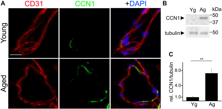
FIGURE 4.
CCN1 was upregulated in the microvessels isolated from the brains of aged mice and in cultured CEC isolated from aged mice compared with young mice. (A) Representative image of microvessels isolated from the brains of young (3–4 m/o) and aged (18–20 m/o) female mice, and stained with antibodies against the endothelial marker CD31, with antibodies against CCN1, and with the nuclear Hoechst dye (DAPI channel). Scale bar, 25 µm. (B) CEC were isolated from young (3-4- m/o) and aged (18–20 m/o) female mice, cultured and maintained until 100% confluence. Cells were collected and lysed and processed for gel electrophoresis and analyzed for Western blotting. (C) Band intensities of CCN1 relative to tubulin. t-test, ** p-value < 0.0019. Data were obtained from four mice per age.

