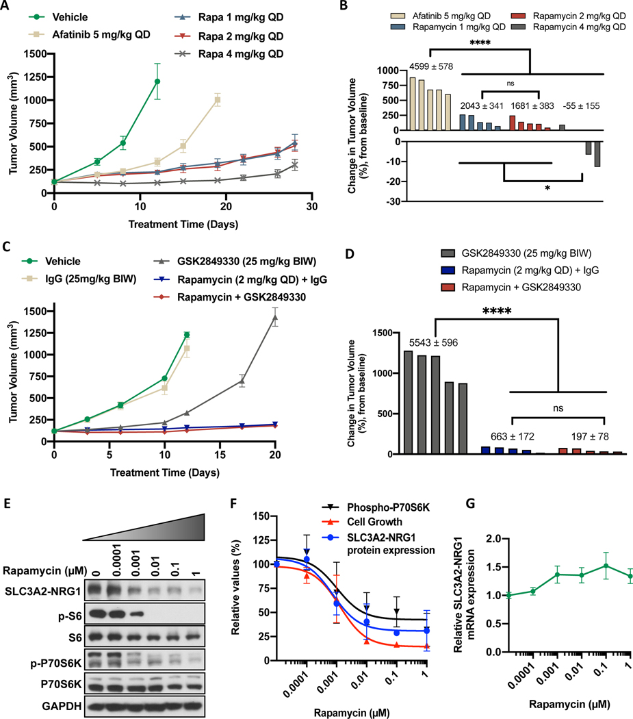
Figure 6. Rapamycin treatment inhibits growth of LUAD-0061AS3 PDX tumors and reduce expression of the SLC3A2-NRG1 fusion.
LUAD-0061AS3 PDX tumors were implanted into a subcutaneous flank of NSG mice and treated as indicated on the graphs. Tumor volume and animal weight were measured twice weekly. Animal weight is given in Supplementary Figure S5. Tumor volume over time (A and C) and change in tumor volume (B and D) are shown. Each group consisted of five animals and data represent the mean ± SE of tumor volumes. Area under the curve (AUC) analysis was used to compare treatment effects and the AUC ± SE values are shown above the bars in B and D for days 19 and 20 respectively. Additional AUC data is provided in Supplementary Figure S5 for days 11 and 12 respectively. E-G. LUAD-0061AS3 cells were treated with the indicated concentrations of rapamycin for 24 h and then either whole-cell extracts were prepared for Western blotting (E). F. SLC3A2-NRG1 levels in comparison to cell viability and phosphorylation of p70S6K as a function of rapamycin concentration. Immunoblots were quantitated by densitometry and results represent the mean ± SD of two independent experiments. G. RNA was isolated from cells treated with rapamycin for 24 h and SLC3A2-NRG1 fusion mRNA level was then determined by qPCR using primers that target SLC3A2 and NRG1. Results are the mean ± SD of three replicate determinations in one experiment. * p < 0.05, **** p < 0.0001, ns: not significant (p > 0.05).

