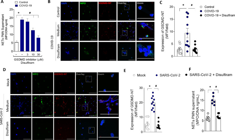
Fig. 2.
GSDMD activation during COVID-19 mediates NETs formation. Human neutrophils were isolated from healthy control (n = 12) and COVID-19 (n = 15) patients. Cells were treated with disulfiram (3, 10, and 30 µM) and cultured for 4 h at 37 °C. A The concentrations of MPO/DNA-NETs in the supernatants were determined using the picogreen test. B Representative immunostaining images for DNA (DAPI, blue), myeloperoxidase (MPO, green), and the GSDMD cleaved fraction (GSDMD-NT, red) are shown. The scale bar indicates 50 μm at 630× magnification. 4 × digital zoom was performed in the inset white square. C GSDMD-NT expression was quantified by MFI per field. D Human neutrophils were isolated from healthy control (n = 6). Cells were treated with disulfiram (30 uM). After 1 h, the cells were incubated with SARS-CoV-2 or Mock (virus control) and cultured for 4 h at 37 °C. Representative immunostaining images for DNA (DAPI, blue), myeloperoxidase (MPO, green), and the GSDMD cleaved fraction (GSDMD-NT, red) are shown. The scale bar indicates 50 μm at 630× fication. 4 × digital zoom was performed in the inset white square. (E) GSDMD-NT expression was quantified by MFI per field. F The concentrations of MPO/DNA-NETs in the supernatants were determined using the picogreen test. The data are expressed as mean ± SEM (*or # p < 0.05; one-way ANOVA followed by Tukey’s test in A, C, E, and F)

