Fig. 6.
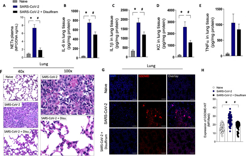
Pharmacological inhibition of GSDMD prevents NET release, lung inflammation, and organ damage in a mouse model of COVID-19. ACE-2 humanized mice were infected with SARS-CoV-2, and after 24 h, mice were treated with disulfiram (50 mg/kg, i.p. 1 × per day, during 5 days) or vehicle. A The MPO/DNA-NET concentration in the plasma was determined 5 days post-SARS- CoV-2 infection. B–E The levels of inflammatory cytokines (IL-6, IL-1β, CXCL-1/KC, and TNF-α) in lung tissue were measured by ELISA 5 days post-SARS-CoV-2 infection. F Representative images of the histological staining of the lung sections performed 5 days post-SARS-CoV-2 infection are shown at 200× magnification and 400× magnification. G Representative confocal analysis of GSDMD-NT and NETs in the lung tissue sample. Immunostaining for DNA (DAPI, blue) and the GSDMD cleaved fraction (GSDMD-NT, red) are shown. The scale bar indicates 50 μm at 630 × magnification. H GSDMD-NT expression was quantified by MFI per field. The data are expressed as means ± SEM (*or # p < 0.05; one-way ANOVA followed by Tukey’s test in A–E and H). The data are representative of at least two independent experiments, each including 5–7 animals per group

