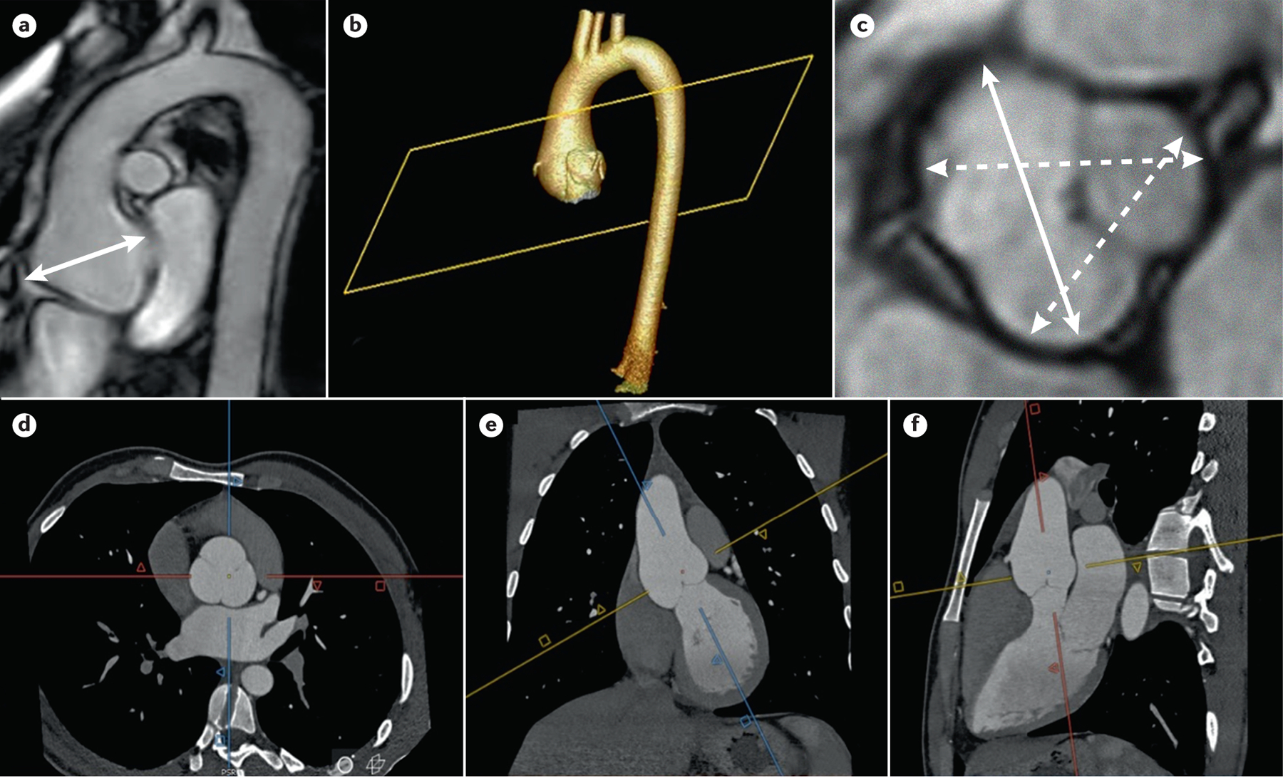
Figure 5. Imaging for thoracic aortic disease in Individuals with MFS.

Imaging for thoracic aortic disease in MFS patients. A) MRI of the thoracic aorta shows an aortic root aneurysm (double arrow). 3D reconstruction (b) of CTA imaging (d, e, and f) of an aortic root aneurysm. The methodology of acquiring double oblique aortic images using the sagittal and coronal images to achieve perpendicularity to the aortic flow results in a corrected true transversal image of the aortic lumen; C) measurement of aortic root aneurysm by MRI using cusp to cusp diameters at end-diastole. Solid double arrow line shows the maximum aortic root diameter.
