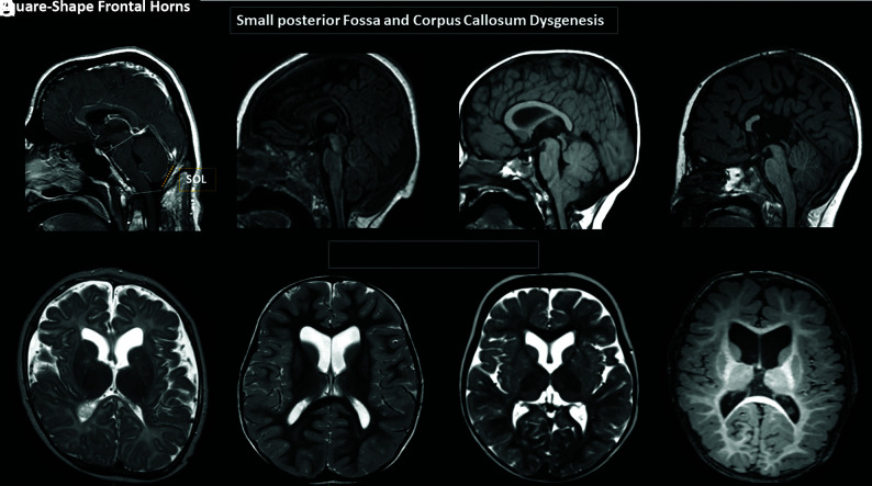
FIG 1.
Brain MR images. A and D, Variable degrees of corpus callosum deformities, particularly involving the body and splenium, noting partial agenesis in D. Deformed morphology of the posterior fossa, with variable degrees of low insertion of the torcula and size reduction of the supraoccipital line (SOL). Note the crowded appearance of the structures in the posterior fossa along with low disposition of the cerebellar tonsils, fitting in the Chiari I deformity criteria in C and D. E–H, Variable degrees of reduced size and/or internal rotational appearance of the head of the caudate nuclei, resulting in an enlarged and squared appearance of the frontal horns.

