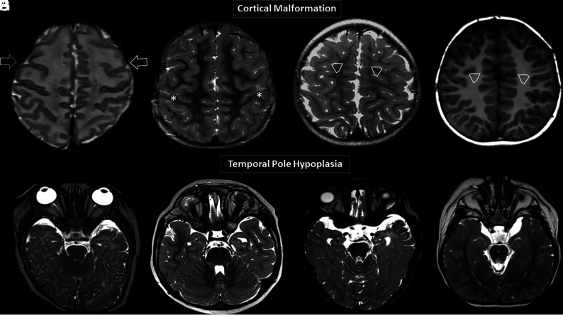
FIG 2.
Brain MR images. Axial T2WI (A–C) and axial T1WI (D) showing 4 different patients with variable degrees of diffuse abnormal orientation and morphology of the gyri and sulci. Note particular abnormal morphology involving both frontal lobes (open arrows, A), a deformed perirolandic region (asterisks, B), and abnormal gyration of the medial frontal lobes in C and D (open arrowheads, C and D). Axial T2WI (E–H) shows 4 different patients with temporal pole hypoplasia.

