Figure 3.

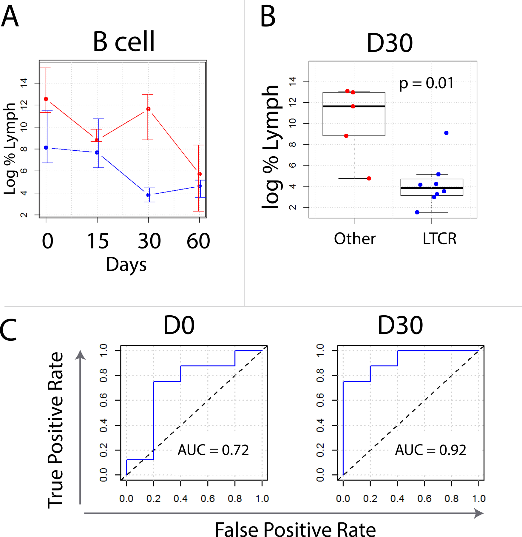
Flow cytometric analysis of B cells in LTCR (blue) versus all other patients (red). (A) Log of median values with error bars indicating 75th and 25th percentiles. Non-LTCR tended to have both more B cells than LTCR as well as less of a Day 30 reduction in B cells following rituximab. Flow gating strategy for these experiments can be found in Supplementary Figure 2. (C) Box whisker plots of quantified B cells on Day 30. When compared to partial responders, complete responders tended to have fewer B cells. (D) Receiver operator characteristic curves constructed for B cells number as a classifier to distinguish LTCR from all others. Area under the curve values are calculated for Days 0 and 30.
