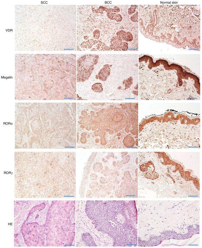
Figure 8.
Immunohistochemical detection of VDR, RORα, RORγ, megalin and hematoxylin and eosin stained sections of human SCC (left column), BCC (middle column) and normal skin (right column) on archival formalin-fixed paraffin-embedded sections. Scale bar=100 µm. Immunohistochemistry was performed on the on archival formalin-fixed paraffin-embedded sections. The methodology for IHC is described in the Materials and methods and with flow chart in Fig. S2. SCC, squamous cell carcinomas; BCC, basal cell carcinomas.

