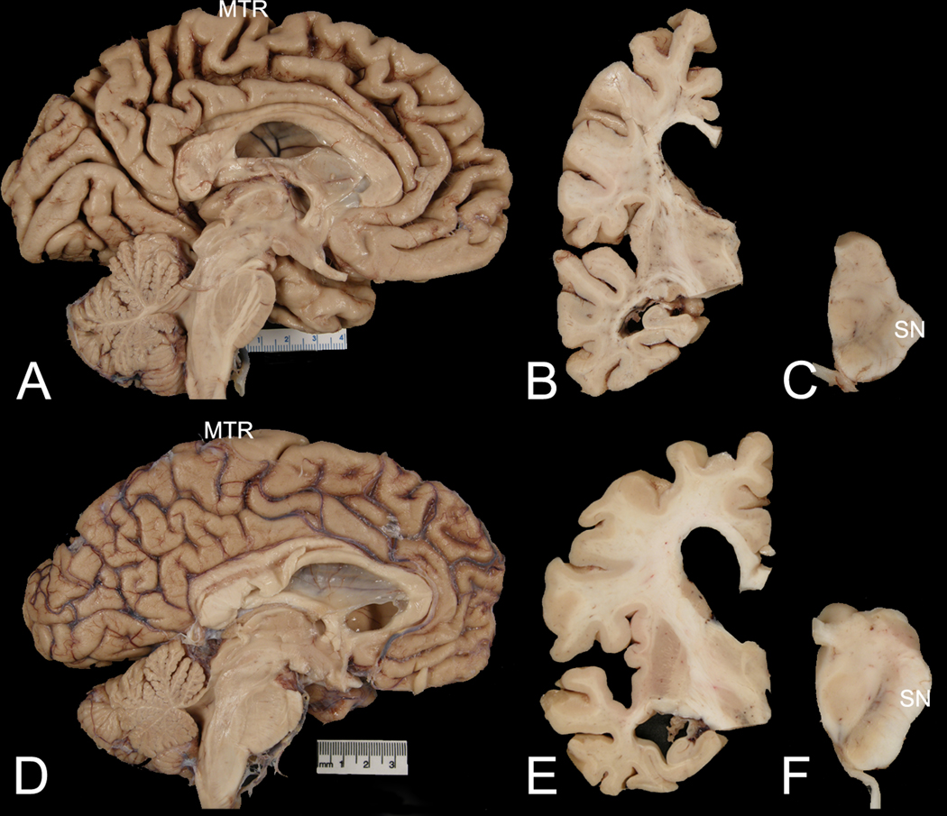
Fig. 2. Macroscopic examples of AD-CBS and AD-AS.

Comparison of representative cases of AD-CBS (A, B, C) and AD-AS (D, E, F). The midsagittal view shows marked medial frontal atrophy, especially in paracentral lobule and peri-Rolandic region (MTR) in AD-CBS (A), but not in AD-AS (D). Coronal sections show ventricular enlargement, with disproportionate enlargement of frontal compared with temporal horns of the lateral ventricle in AD-CBS (B) compared with AD-AS (E). Transverse sections of the midbrain show decreased neuromelanin pigment in the lateral substantia nigra (SN) in AD-CBS (C) compared with AD-AS (F).
