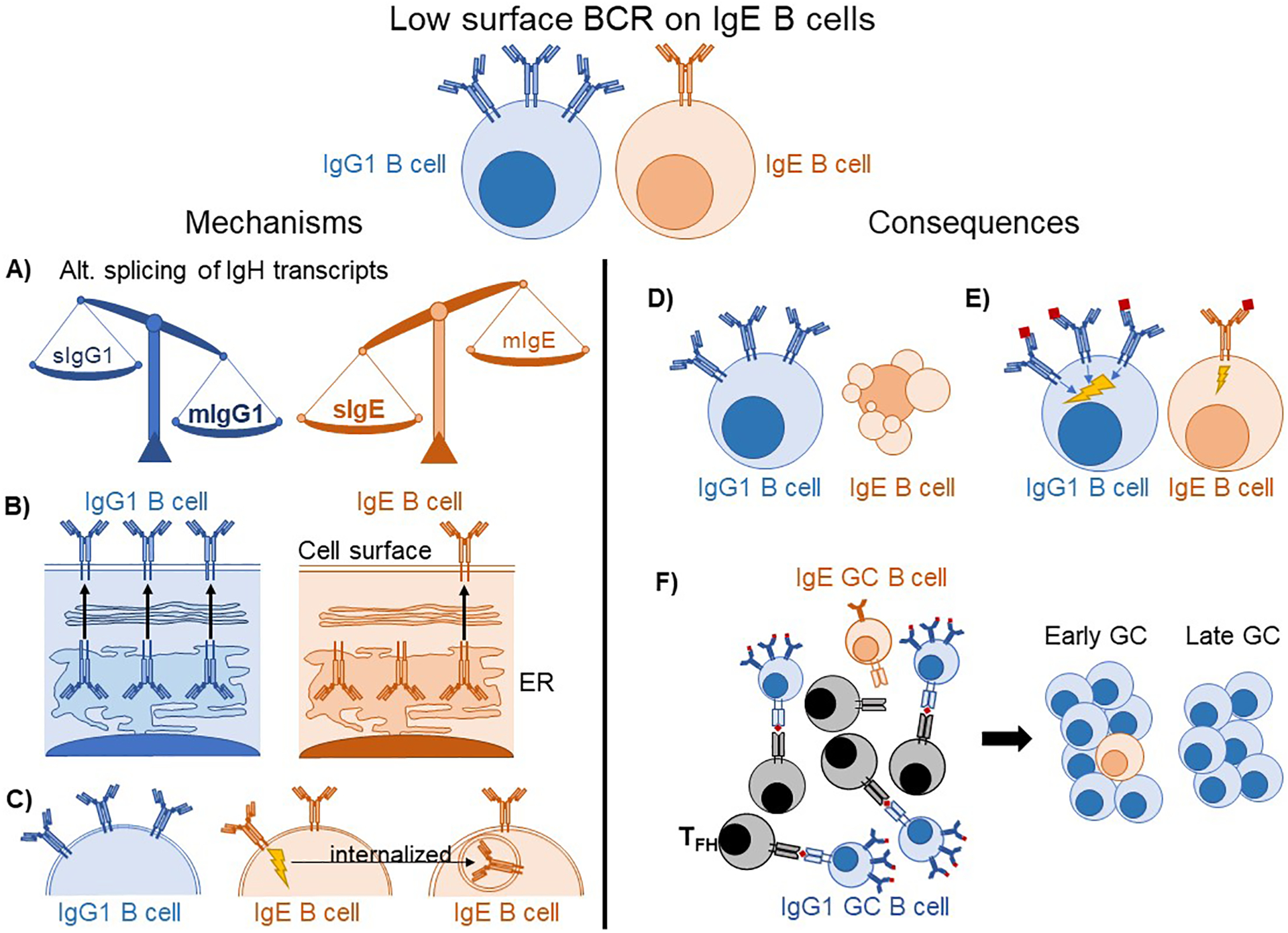
Figure 1: Proposed mechanisms and consequences of low surface BCR expression on IgE B cells.

(A–C) Potential mechanisms explaining low IgE BCR surface expression. (A) IgG1 B cells favor the mIg spliceoform of IgH, whereas IgE B cells favor the sIg spliceoform, resulting in fewer mIgE transcripts available for the translation of IgE BCR molecules. (B) The IgE BCR is retained in the ER after translation rather than being exported to the cell surface. (C) Antigen-independent signaling of the IgE BCR leads to its internalization, reducing surface BCR levels. (D–F) Putative consequences of low BCR expression on IgE B cells. (D) IgE B cells undergo apoptosis due to insufficient BCR expression. (E) IgE B cells have weaker antigen-dependent BCR signaling than IgG1 B cells. (F) IgE GC B cells are less able to capture and present antigens to GC TFH, causing them to be progressively out-competed over time.
