Figure 2.

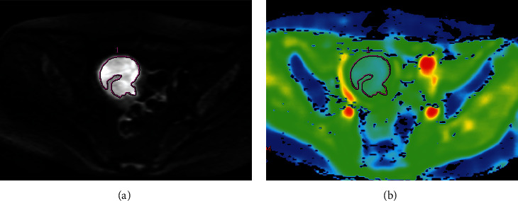
DWI image of a 57-year-old patient with EC of stage IB. (a) ROI was outlined at the largest tumor cross section on DWI (b = 1000 s/mm2). (b) ADC pseudocolor image showed that the ADC was 0.966 × 10⁻³ mm2/s.

DWI image of a 57-year-old patient with EC of stage IB. (a) ROI was outlined at the largest tumor cross section on DWI (b = 1000 s/mm2). (b) ADC pseudocolor image showed that the ADC was 0.966 × 10⁻³ mm2/s.