Figure 1.

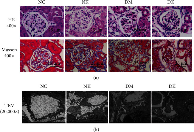
(a) Pathological examination of renal tissues of mice in each group by light microscopy (400x). (b) The ultrastructure of renal tissue was observed by transmission electron microscope (20,000x).

(a) Pathological examination of renal tissues of mice in each group by light microscopy (400x). (b) The ultrastructure of renal tissue was observed by transmission electron microscope (20,000x).