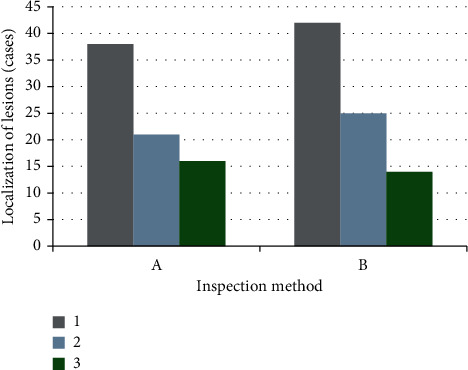
Figure 6.

Comparison of DWI combined with TCD, CT, and MRI in locating lesions. A referred to the results of DWI combined with TCD; B showed the results of CT and MRI. 1, 2, and 3 referred to the number of patients with infarction of the internal carotid artery system, infarction of the vertebral base arterial system, and the ischemic infarction of the marginal zone between adjacent blood vessels, respectively.
