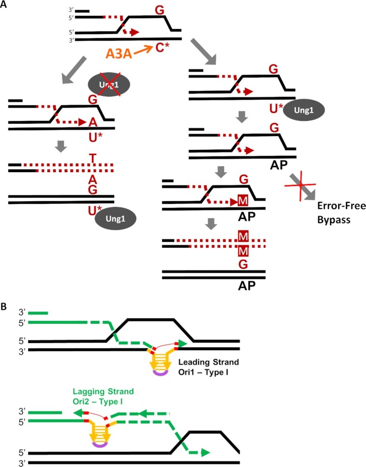
Figure 6.

(A) Cytidine (C*) in the ssDNA of D-BTS is susceptible to A3A-induced deamination producing dU lesions (U*). The fate of dU lesions follows one of two possible paths. In the first (left), the Ung1 enzyme is unable to excise the dU base, leaving it in the template where it is encountered by leading strand synthesis. An A base is placed across from the dU lesion, incorporating it into the nascent strand where it is paired with a T base during lagging strand synthesis. Ung1 may later repair the template strand dU lesion after BIR has proceeded beyond it, but the mutation will stay in the newly synthesized strand. In the second path (right), Ung1 is able to access the dU lesion created by A3A and excise it, leaving an AP-site. This AP-site does not (or rarely) triggers error-free bypass pathways. Instead, a base placed across from the AP site during leading-strand synthesis often results in incorporation of the wrong base into the nascent strand, resulting in a mutation (M). (B) Schematics of Type I InsH deletion following hairpin formation during leading or lagging strand synthesis of BIR. Directions of leading and lagging strand synthesis are defined by the known direction of BIR synthesis for BIR in our system. Microhomologies at InsH deletion breakpoints are indicated in red.
