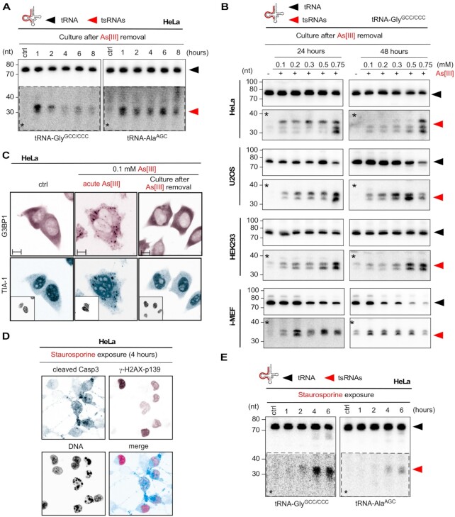
Figure 5.
(A) Northern blotting of total RNA (1.5 μg) extracted from HeLa cells after time-limited exposure (1 h) to As[III] (0.5 mM), followed by culturing cells for the indicated times (1, 2, 4, 6, 8 h) after the removal of As[III], using probes against the 5′ ends of tRNA-GlyGCC/CCC and tRNA-AlaAGC. Black arrowhead: mature tRNAs; red arrowhead: tsRNAs; asterisks: digitally enhanced against parental tRNA signals. (B) Northern blotting of total RNA (3 μg) extracted from HeLa, U2OS, HEK293 cells and i-MEF, which had been exposed (1 h) to increasing molarities of As[III], followed by culturing cells for 24 or 48 h after the removal of As[III] using a probe against the 5′ end of tRNA-GlyGCC/CCC. Black arrowhead: mature tRNAs; red arrowhead: tsRNAs; asterisks: digitally enhanced against parental tRNA signals. (C) Indirect immunofluorescence image of HeLa cells after time-limited exposure (1 h) to As[III] (0.5 mM), followed by culturing cells for 24 h after the removal of As[III], using antibodies against G3BP1 (magenta) and TIA-1 (cyan). Individual insets: DNA (black). Scale bar 10 μm. (D) Indirect immunofluorescence image of HeLa cells after exposure (4 h) to staurosporine (1 μM), using antibodies against cleaved caspase 3 (cyan) as indicator of apoptotic cells and phosphorylated γ-H2AX (magenta) as indicator of DNA damage. DNA: black. (E) Northern blotting of total RNA (1.5 μg) extracted from HeLa cells, which had been exposed to staurosporine (1 μM) for the indicated times (1, 2, 4, 6 h) using probes against the 5′ ends of tRNA-GlyGCC/CCC and tRNA-AlaAGC. Black arrowhead: mature tRNAs; red arrowhead: tsRNAs; asterisks: digitally enhanced against parental tRNA signals.

