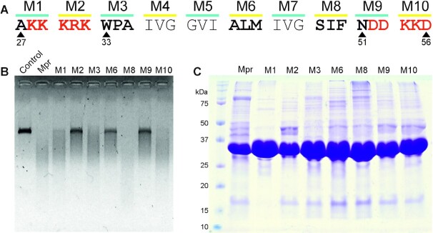
Figure 6.
Nuclease activity of Mpr with alanine-substituted mutants. (A) Schematic representation of the TM region of Mpr; charged residues flanking the TM region are depicted in red. Residues mutated to alanine are highlighted in bold; respective positions of residues are mentioned. (B) Agarose gel image showing nuclease activity of Mpr and its mutants is presented; dialysis buffer is used as control. (C) Coomassie-stained SDS-PAGE gel image of purified Mpr and its mutants is presented; first lane corresponds to protein ladder with size of various protein bands marked for reference.

