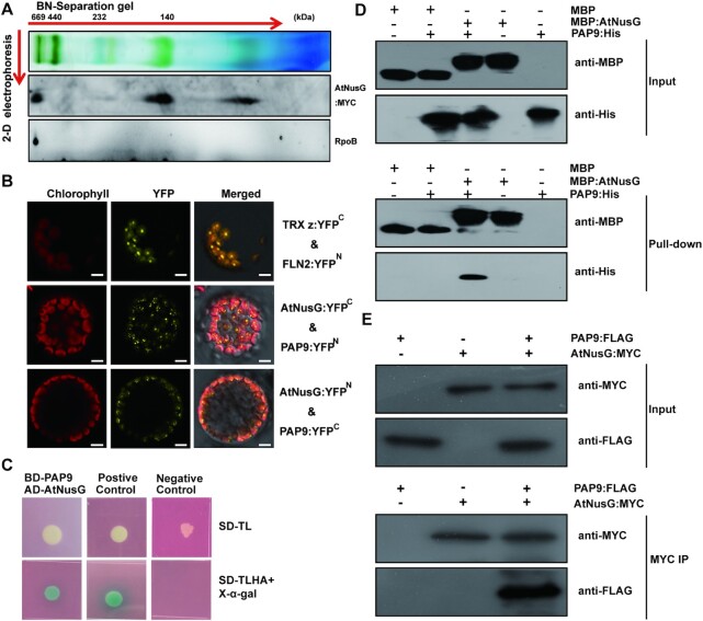
Figure 2.
AtNusG is associated with the chloroplast PEP complex by interacting with PAP9 in Arabidopsis. (A) Immunoblotting detection of AtNusG and RpoB by two-dimensional gel electrophoresis. Thylakoid membrane proteins from 3-week-old AtNusG:MYC seedlings were fractionated by BN-PAGE in the first dimension and by SDS–PAGE in the second dimension. The approximate molecular masses of the labelled protein complexes are indicated above. (B) Visualization of protein interactions between AtNusG and PAP9, the essential component of the PEP complex, through the BiFC assay. YFP indicates the fluorescence signals from Arabidopsis protoplasts transiently expressing constructs encoding the fusion proteins. Merge indicates an overlap of the YFP fluorescence and chlorophyll autofluorescence images. The combination of TRX z:YFPC and FLN2:YFPN, which were used as a positive control, was cotransformed into Arabidopsis protoplasts. Bars = 10 μm. At least two independent BiFC assays for each combination were performed. (C) Interaction between PAP9 and AtNusG proteins in yeast two-hybrid assays. Yeast cells containing the combination of BD-PAP9 and AD-AtNusG vectors were grown on selection medium, SD-Leu-Trp and SD-Leu-Trp-His-Ade with X-α-Gal. Yeast cells containing the combination of pGBKT7-53 and pGADT7-T were used as a positive control, while yeast cells containing the combination of pGBKT7-Lam and pGADT7-T were used as a negative control. (D) In vitro pull-down assays between MBP-AtNusG and PAP9-His. Representative immunoblot results of input samples (input) and samples pulled down with anti-MBP antibody are shown. (E) Coimmunoprecipitation assay revealing the in vivo interaction between AtNusG and PAP9. The combinations of AtNusG:MYC and PAP9:FLAG indicated above each blot were transiently expressed in N. benthamiana. Proteins detected by immunoblotting are indicated on the right. PAP9:FLAG coimmunoprecipitated with AtNusG:MYC when the anti-MYC antibody (MYC IP) was used, and immunoblotting was performed with the anti-FLAG antibody.

