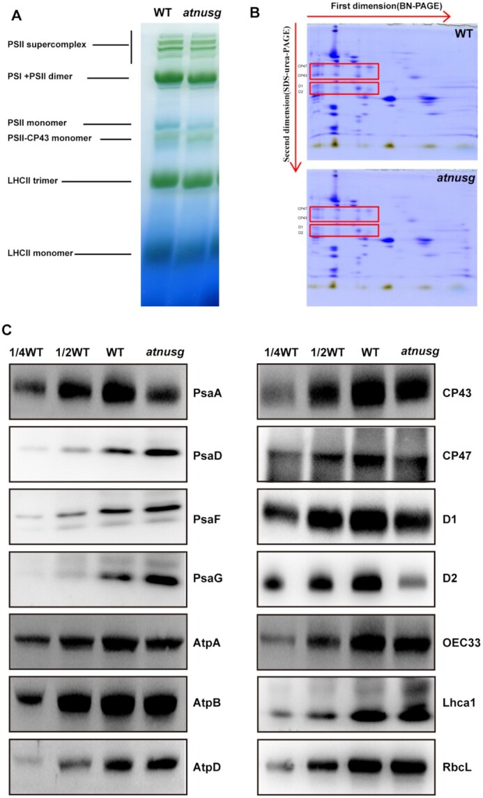
Figure 5.

Analysis of photosynthetic supercomplexes from the wild type and atnusg mutant under normal growth conditions. (A) BN-gel analysis of thylakoid membrane protein complexes in the wild type (WT) and atnusg mutant. Representative unstained BN-PAGE gel electrophoresis is shown. Thylakoid membranes from the wild type (WT) and atnusg mutant were solubilized with 1% DM and separated by native PAGE. A sample with an equal amount of chlorophyll (18 μg) was loaded in each lane. The bands for supercomplexes are indicated on the left. (B) 2D, BN/SDS–urea-PAGE electrophoresis analysis of the thylakoid membrane complexes. Thylakoid membrane complexes were separated by BN-PAGE and further subjected to 2D SDS–PAGE. The gels were stained with Coomassie Brilliant Blue. The locations of these plastid-encoded core proteins of PSII (CP43, CP47, D1 and D2) are marked by red boxes on the gels. (C) Immunoblot analysis of the photosynthetic proteins from the wild type (WT) and atnusg mutant. Samples were prepared from emerging leaves of 14-day-old seedlings and then separated by SDS–PAGE.
