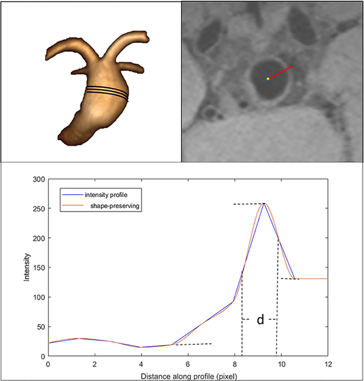
Figure 1.

Algorithm of aneurysm wall thickness calculation. Top left: fusiform basilar aneurysm. Top right: a line is drawn from the center of the aneurysm to the outside wall on a pre-contrast image. Bottom: signal intensity profile. The distance between two half peak points d×0.4 mm was defined as the thickness at that point.
