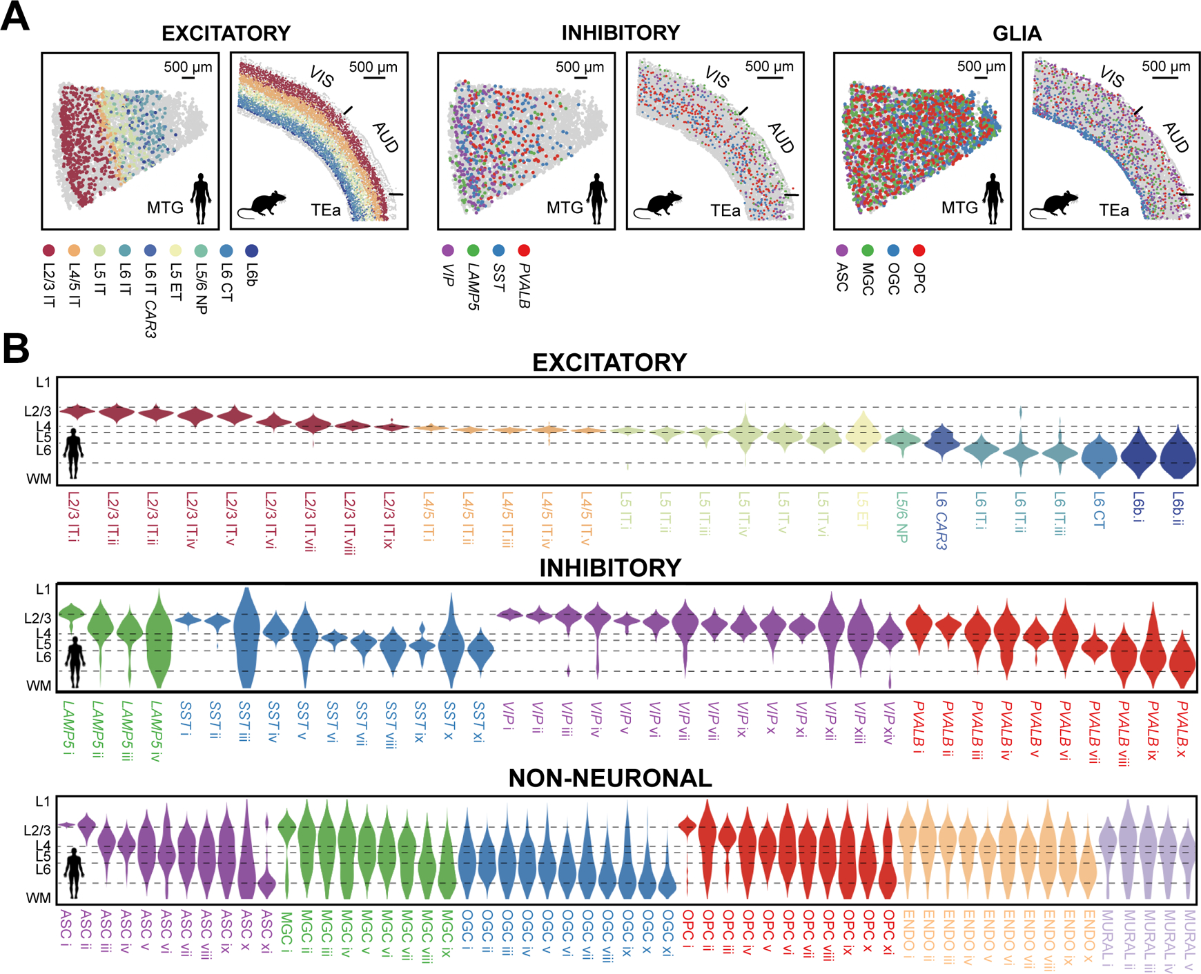
Fig. 2. Laminar organization of cell types in the human and mouse cortex.

(A) Spatial maps of subclasses of excitatory neurons, inhibitory neurons, and glial cells determined by MERFISH in a human MTG slice and a mouse slice containing VIS, AUD and TEa. Indicated subclasses are shown in colors and other cells are in grey. (B) Cortical-depth distribution of excitatory (top), inhibitory (middle) and non-neuronal (bottom) clusters in the human MTG. The dashed grey lines mark the approximate layer boundaries. WM: white matter.
