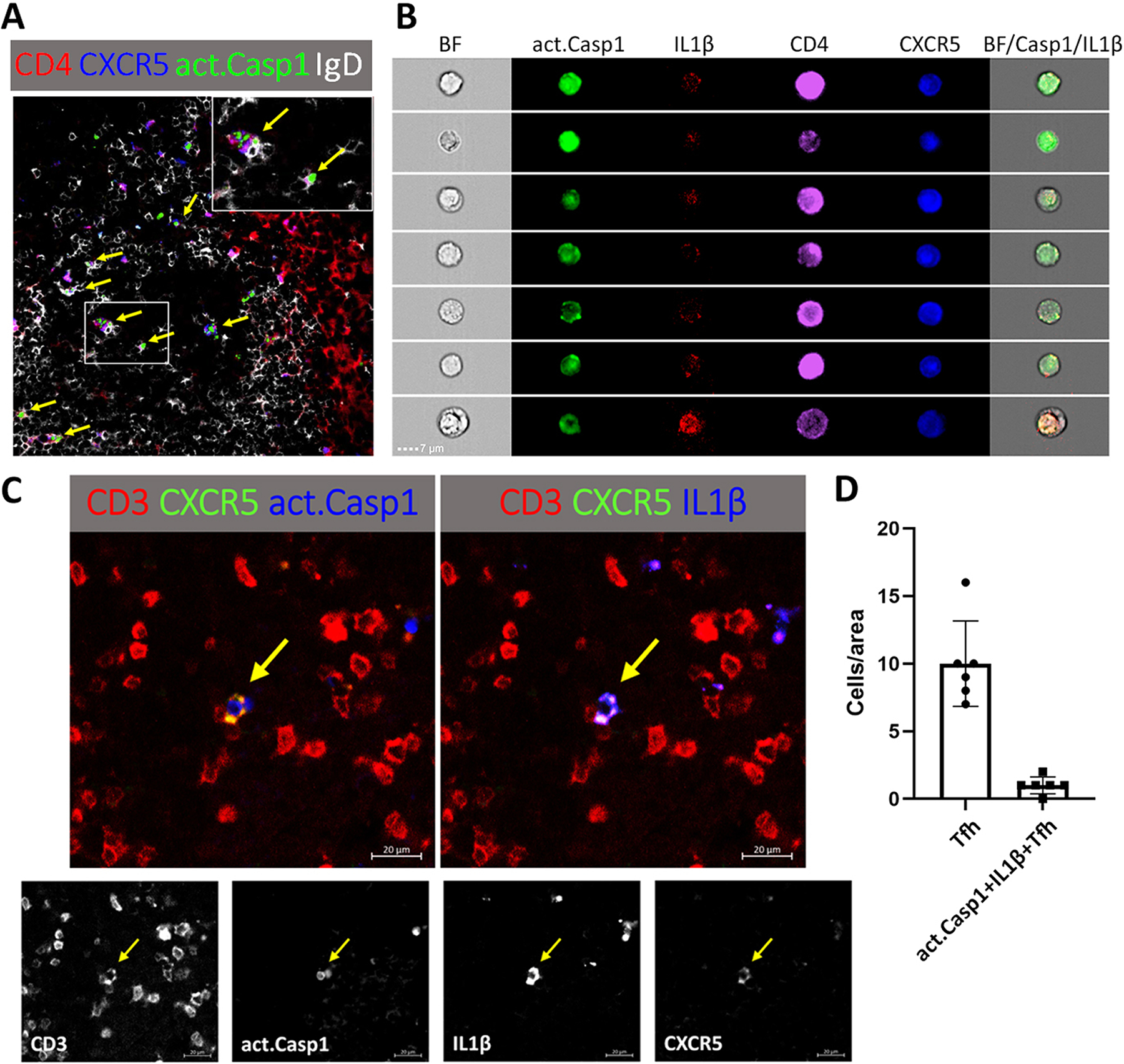
Figure 1. CXCR5+ CD4+ or CD3+ Tfh cells in the lymph node of a B6 mouse five days after the immunization with NP33-CGG with alum in the hind footpads express act. caspase-1+ and IL-1β.

(A) Many of the act. caspase-1+ Tfh cells in the interface between T cell B cell zones and in the dark zone of the GC are identified by arrows. The inset shows interaction between act. caspase-1+ Tfh cells with IgD+ B cells. (B) CD4+CXCR5+ T cells as seen by the ImageStreamX MKII imaging flow cytometer with act. caspase-1 stained green, IL-1β red, CD4 purple and CXCR5 blue. (C) A CD3+CXCR5+ T cell stained for act. caspase-1 and IL-1β. (D) CD3+CXCR5+Tfh and caspase-1+ IL1β+ Tfh were counted in each 300 μm × 300 μm area.
