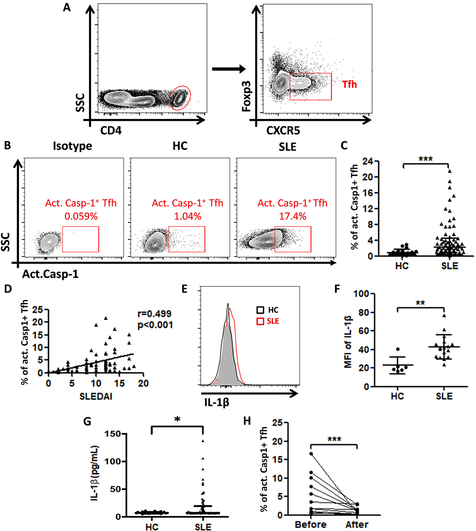
Figure 7. Increased activation of NLRP3 inflammasome in circulating Tfh cells of SLE patients are correlated with SLEDAI and response to therapy.

(A) Gating strategy to identify circulating Tfh cells (CD4+CXCR5+Foxp3−) in human blood. (B) Contour plots of act. caspase-1+ Tfh cells from a representative healthy control and a SLE patient. (C) Percentage of act.caspase-1+ Tfh cells among total Tfh cells in SLE patients (n=71) and healthy controls (n=25). (D) Correlation between the SLEDAI with the percentage of act. caspase-1+ Tfh cells in SLE patients. (E) Flow-cytometric histograms of IL-1β expression in Tfh cells of a representative healthy control (Black line) and a SLE patient. (Red line). (F) MFI of IL-1β on Tfh cells of HCs (n=6) and SLE patients (n=17). (G) Serum IL-1β levels of HCs (n=25) were compared with those in SLE patients (n=71). (H) Percentage of act. caspase-1+ Tfh cells before and after treatment (n=11). *p<0.05, **p<0.01 and ***p<0.001.
