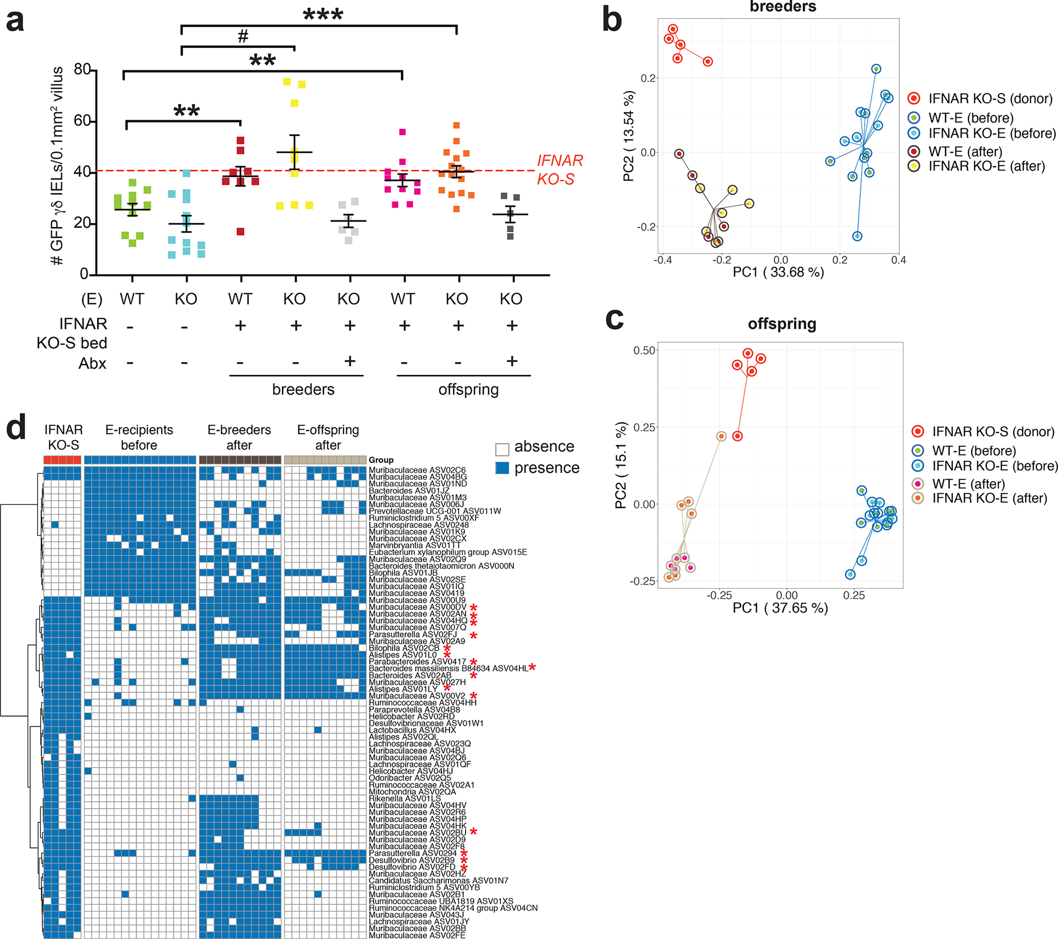
Figure 2. Horizontal transfer of the microbiota is necessary and sufficient to induce the γδ IEL hyperproliferative phenotype.

a Morphometric analysis of the number of GFP+ γδ T cells in untreated WT-E, IFNAR KO-E mice; WT-E or IFNAR KO-E breeders and offspring following IFNAR KO-S bedding transfer in the presence or absence of antibiotic (Abx) treatment. Dashed line indicates the number of γδ IELs in IFNAR KO-S donor mice. n=5–15. Principal coordinates analysis of 16S rRNA sequencing of fecal microbiota from donor and recipient breeders b and offspring c n=4–6. d Binary heatmap of 69 ASVs shared in recipient mice (breeders and offspring) following horizontal transfer of microbiota. The red asterisk highlights the 15 ASVs which had significantly lower prevalence in pre-transfer breeders compared with donors, post-transfer breeders and post-transfer breeders’ offspring. Each data point represents an individual mouse. Data represent mean (±SEM). Statistical analysis: a: one-way ANOVA with Dunnett’s post hoc test **P<0.01, *** P<0.001, # P<0.0001; d: Fisher’s exact test *P<0.05
