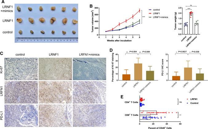
Fig. 6.
LRFN1 promotes ccRCC growth and immune-infiltrating TIME on a xenograft model. A The representative images of three groups (control, LRFN1 overexpression plasmid, LRFN1 plus miR-187-3p mimics) on the xenograft model. B miR-187-3p rescues LRFN1 promoted A498 cell volume and weight in vivo. C, D The IHC staining was performed to assess Ki-67 staining, LRFN1 expression and PD-L1 expression of three groups. E The Flow cytometry assay was performed to reveal active CD4+ and CD8+ T cells in tumor tissues between high and low LRFN1 expression groups. *P < 0.05; **P < 0.01; ***P < 0.001

