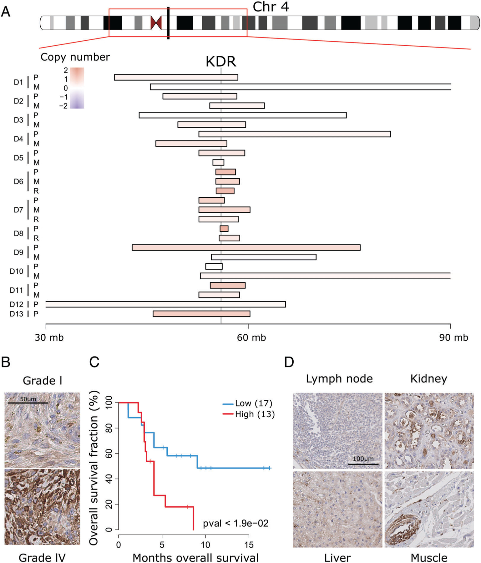
Figure 5.

KDR is recurrently amplified in osteosarcoma and associates with poor outcome. (A) Relative copy number status of different chromosome four segments, including the KDR genomic locus, showing recurrent copy number gains (red) across the tumor cohort. (B) Representative examples of KDR IHC of Grade I and IV osteosarcomas. (C) Kaplan–Meier curves of patients with high (H-score > 15) compared to low (H-score ≤ 15) KDR (p < 0.05, log rank test). (D) KDR IHC of the indicated normal organ tissue sections. Only vascular endothelial cells show appreciable immunoreactivity, as shown in the inserts.
