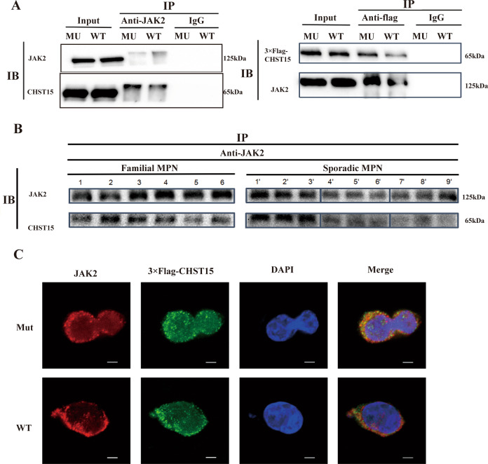
Fig. 6. JAK2/CHST15 complex formation.
A Coimmunoprecipitation between JAK2 and CHST15 on HELcells harbouring CHET15 mutation (CHST15R456fs) or wild-type CHST15. left panel:anti-JAK2 antibody; right panel:anti- Flag-CHST15 antibody. Input, non-immunoprecipitated cell lysates; IgG, control IP with isotype antibody. B Coimmunoprecipitation between JAK2 and CHST15 on primary PBMNCs of patients with familial MPN (left panel: 1, 2, Familial 1. PMF: 3, 4. Familial 3. ET; 5,6. Familial 2, PMF.) or sporadic MPN (right panel: 1’, 2’, 3’: sporadic MPN with JAK2V617F; 4’, 5’, 6’: MPN with CALR mutation; 7’,8’,9’: MPN without mutation- triple negative). C Immunofluorescent colocalization of FLAG-tagged-CHST15 and JAK2. 4′,6-diamidino-2-phenylindole (DAPI) wasused for nuclear staining. The results show that both JAK2 and CHST15 are mainly found on cytomembrane and cytoplasm, and form co-precipitatation with granular shape between CHET15 and JAK2, suggesting exists in physical interaction both JAK2 and CHST15 (wild type) especially CHST15R456fs mutant.

