Figure 1.
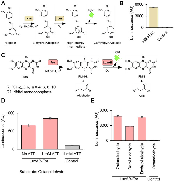
Characterization of H3H-Lux and LuxAB-Fre luciferase systems in TXTL. (A) Schematic of H3H-Luz luciferase reaction. Hispidin is converted to 3-hydroxyhispidin by hispidin-3-hydroxylase (H3H) and 3-hydroxyhispidin is oxidized and converted into a high energy intermediate by the luciferase (Luz). This intermediate decays into caffeylpyruvic acid with light emission. (B) The H3H-Luz luminescence measurement. H3H and Luz were expressed in TXTL. The luminescence was measured right after adding NADPH and hisipidin into the TXTL. (C) Schematic of LuxAB-Fre luciferase reaction. Oxidized flavin mononucleotide (FMN) is reduced into reduced flavin mononucleotide (FMNH2) by NAD(P)H-flavin reductase (Fre). The luciferase (LuxAB) converts FMNH2 and long-chain aldehydes into FMN and the corresponding long-chain acids with light emission. (E) ATP supplementation increased the light emission of LuxAB-Fre. Octanaldehyde was added as the substrate. (D) The LuxAB-Fre luminescence measurement with different long-chain fatty aldehydes. LuxA, LuxB and Fre were expressed in TXTL. The luminescence was measured right after adding FMN, NADPH, ATP, and substrates (octanaldehyde, decyl aldehyde, and dodecyl aldehyde.) NADPH, nicotinamide adenine dinucleotide phosphate; ATP, adenosine triphosphate; Control, reaction without enzyme expression. The graphs show means with error bars that signify SEM (n = 3).

