Figure 2.
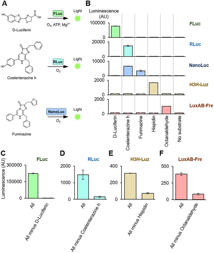
Characterization of substrate specificities. (A) Schematic image of firefly luciferase (FLuc), renilla luciferase (RLuc), and NanoLuc luciferase (NanoLuc) reactions. FLuc oxidizes D-luciferin with ATP and Mg+ to produce light. RLuc and NanoLuc oxidize coelenterazine h and furimazine, respectively, with ATP to produce light. (B) Luminescence measurement for substrate specificity assay for 5 luciferases. The luciferases (FLuc, RLuc, NanoLuc, H3H-Luz, and LuxAB-Fre) were expressed in TXTL. Then, the individual substrates (D-luciferin, coelenterazine h, furimazine, hispidin, and octanaldehyde) with corresponding co-factors were added to the reaction and measured its light emission without emission filters. Substrate concentrations were 10 μM, except 1 mM for octanaldehyde. (C–F) The substrate multiplexing assay. The substrate mixtures were prepared as “All” (D-luciferin, Coelenterazine h, hispidin, octanaldehyde, Mg+, ATP, NADPH, FMN) or “All minus one” that contains all except one that a substrate is supposed to react with a tested luciferase. The assay was performed by mixing substrates with TXTL expressing (C) FLuc, (D) RLuc, (E) H3H-Luz, or (F) LuxAB-Fre, and the luminescence was measured without emission filters. ATP, adenosine triphosphate. The graphs show means with error bars that signify SEM (n = 3).

