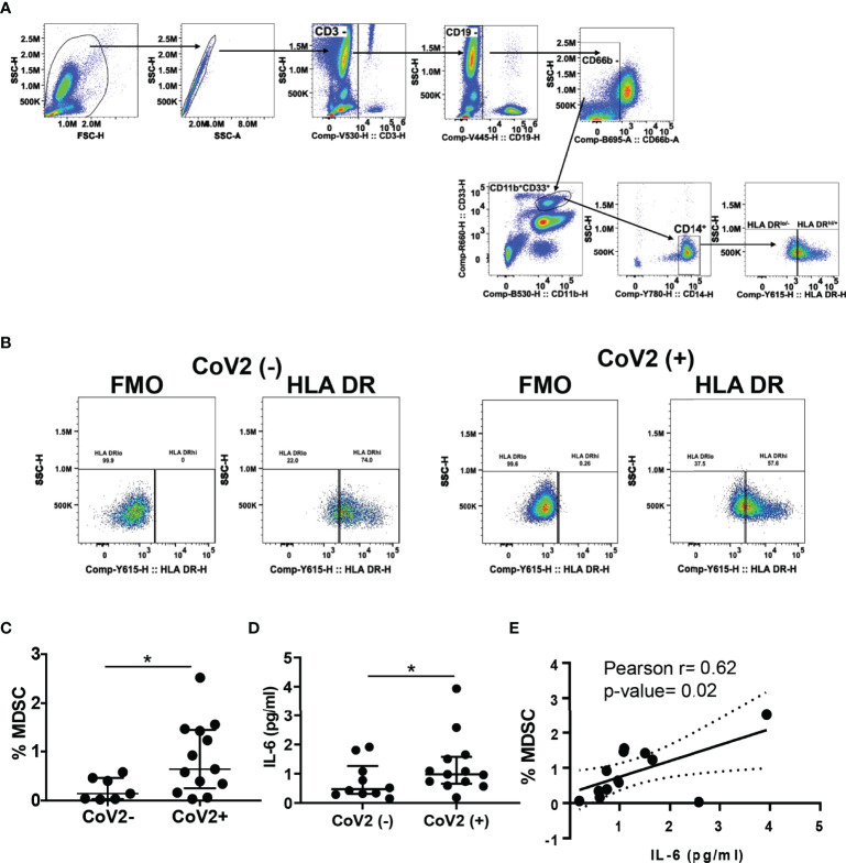
Figure 1.
M-MDSC expansion in CoV2+ individuals is dependent on IL-6: (A–C) Heparinized blood obtained from CoV2- and CoV2+ individuals was stained with anti-CD3, -CD19, -CD11b, -CD33, -CD14, CD66b, -HLA DR antibodies, cells were analyzed as CD3-CD19-CD66b-CD11b+CD33+CD14+HLA DR-/lo by flow cytometry. (A) Gating strategy for MDSC is shown (B) A representative dot plot with HLA DR-/lo region from CoV2(-)and CoV2(+)is shown. (C) Percentages of MDSC are shown. (D) The quantity of cytokine IL-6 in the plasma of CoV2- and CoV2+ individuals was measured by ELISA, as in Methods. (E) Plasma IL-6 quantity was correlated with the circulating frequency of M-MDSC in CoV2+ individuals. (C, D) Each dot in the plots depicts data of each individual donor, the plots include observations from 25th to 75th percentile. The horizontal line represents the median value. (D) Each dot in the plot depicts data of each individual donor; black solid and dotted lines, model-estimated values, and their 95% confidence intervals. *p < 0.05.

