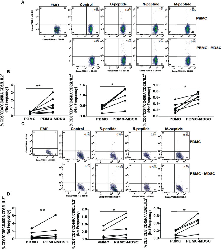
Figure 3.
M-MDSC regulates CoV2 antigen-specific IL-2 production: Freshly isolated PBMC from CoV2+ individuals were stained with anti-CD14 and -HLA DR antibodies; CD14+HLA DR-/lo M-MDSC were depleted from PBMC by flow cytometry. Whole PBMC (PBMC) and MDSC depleted PBMC (PBMC-MDSC) were cultured in the absence or presence of peptide pools of S-, N-, and M- antigens of CoV2 for 20-24 hours. Cells were stained with anti-CD3, -CD4, -CD8, -CD45RA, -CD62L, IL-2 antibodies, and LIVE/DEAD fixable stain. (A, B) Percentages of CD3+CD4+CD45RA-CD62L-IL-2+ cells was determined. (C, D) Percentages of CD3+CD8+CD45RA-CD62L-IL-2+ cells was determined. (A, C) Representative dot plots showing CD4+IL-2+ gated on Live CD3+CD4+CD45RA-CD62L- (A), and CD8+IL-2+ gated on Live CD3+CD8+CD45RA-CD62L- (C) are shown. (B, D) Each dot represents an individual donor. *p < 0.05, **p < 0.005, FMO, Fluorescence minus one.

