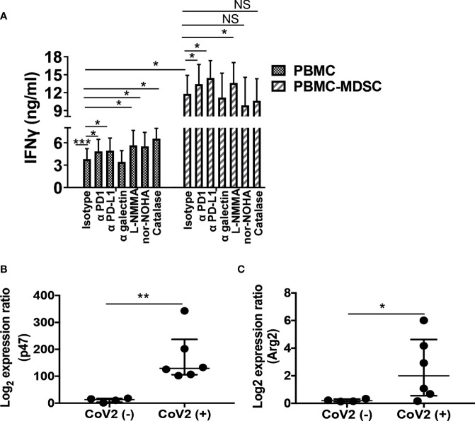
Figure 5.
M-MDSC mediated T cell inhibition is dependent on Arg and ROS: Freshly isolated PBMC from CoV2- and CoV2+ individuals were stained with anti-CD14 and -HLA DR antibodies; CD14+HLA DR-/lo M-MDSC were depleted from PBMC by flow cytometry. (A) Whole PBMC (PBMC) and MDSC depleted PBMC (PBMC-MDSC) of CoV2+ individuals were cultured in the absence or presence of the peptide pool of S-antigen in presence of blocking anti-PD1, -PD-L1, -galectin antibodies or isotype-matched control antibody, or in presence of iNOS inhibitor (L-NMMA), arginase inhibitor (nor-NOHA) or ROS inhibitor (Catalase). Supernatants were collected at 48-72 hours and the quantity of IFNγ was measured by ELISA. (B, C) RNA was isolated from sorted CD14+HLA DR-/lo MDSC using Trizol. The expression of (B) p47phox (p47) and (C) Arg2 was determined by qRT PCR as detailed in Methods. For (A) Mean values +/- SEM are shown from 4 separate donors; (B, C) each dot in the plots depicts data of each individual donor, the plots include observations from 25th to 75th percentile. The horizontal line represents the median value. *p < 0.05, **p < 0.005; ***p < 0.0005; NS, Non-significant.

