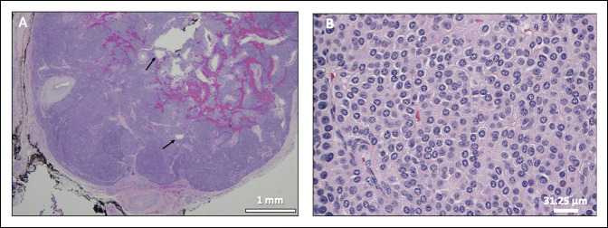
Figure 4.
A, Image showing a circumscribed glomus tumor with a fibrous capsule, dilated vessels (arrows), hemorrhage, and nests/sheets of tumor cells separated by fibrous bands. B, Image showing uniform small round cells with distinct cell borders, centrally placed round-to-oval nuclei with relatively coarse chromatin, and granular eosinophilic cytoplasm. Hematoxylin and eosin staining.

