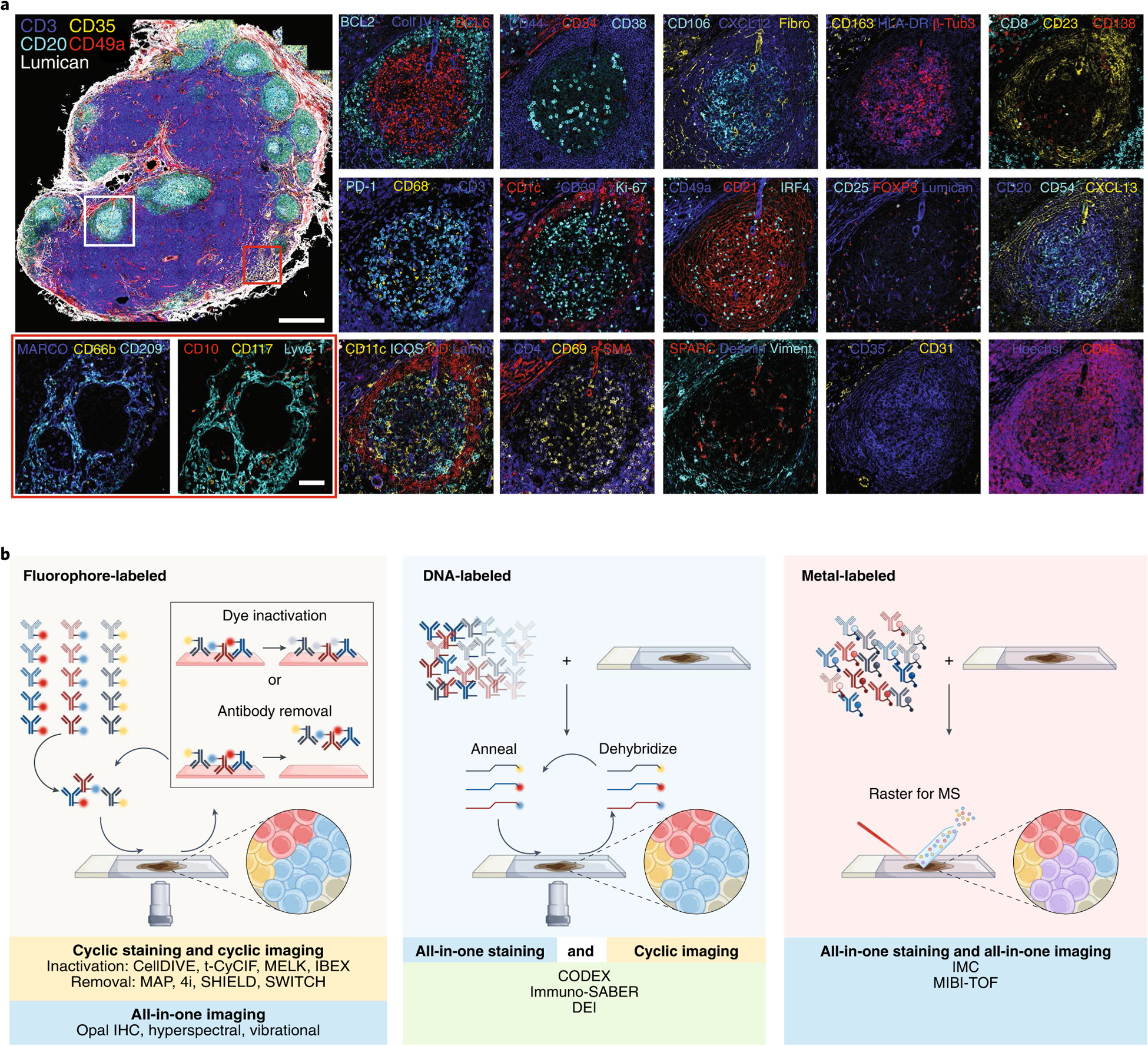
Fig. 1 |. obtaining high-content imaging data using a wide range of multiplexed antibody-based imaging platforms.

a, Fifty-plex confocal images of a human mesenteric lymph node obtained by the IBEX method. Two to four marker overlays for two regions (germinal center, white rectangle and medullary cords, red rectangle) are shown in higher zoom. Scale bars, 500 and 100 μm, for the overview and zoom-in images, respectively. β-Tubulin 3 (β-Tub3), collagen IV (Coll IV), fibronectin (Fibro), laminin (Lamin), and vimentin (Viment) (original lymph node dataset from Radtke et al.21). b, Graphical representation of the main approaches for multiplexed antibody-based imaging. Antibodies are commonly labeled with metals, fluorophores, or DNA oligonucleotides for complementary binding of fluorescently tagged DNA probes.
