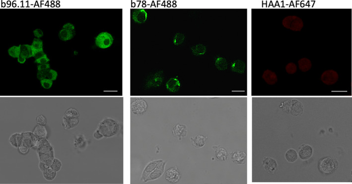
FIGURE 1.

Confocal imaging of dispersed islet cells incubated with mAbs b96.11, b78 and HAA1 conjugated to the indicated fluorochromes. Dispersed islet cells were grown on coverslips at 50‐100,000 cells/well, fixed and permeabilized, and incubated with 2μg/ml of antibody for 2 h. Top row of panels: fluorescent images. Lower row of panels: bright field images of the respective cells shown in the above panel. Roughly 100 cells were analyzed for each image with ~60% showing positive staining with GAD65 mAb. The images are typical individual cells found within a single set of dispersed islets and are representative of similar analyses of six independent rat islet isolations. Scale bar is 10 microns in length
