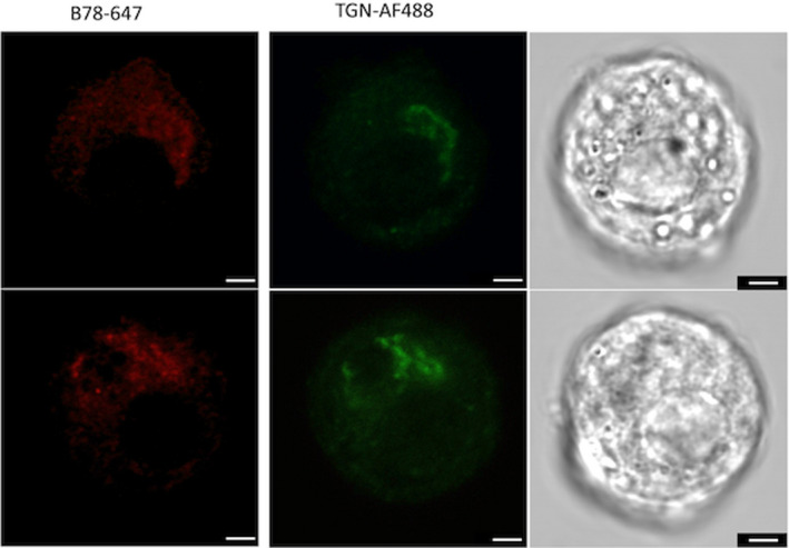
FIGURE 3.

Confocal imaging of dispersed islets incubated with GAD65 mAb b78‐AF647 and TGN‐AF488. Bright field images of the respective cells are shown in the right panels. Dispersed islet cells were grown on coverslips at 50‐100,000 cells/well, fixed and permeabilized, and incubated with 2μg/ml of antibody for 2 h. Roughly 100 cells were analyzed. The images are typical of individual cells found within a single set of dispersed islets and are representative of similar analyses of three independent rat islet isolations. Scale bar is 2.5 microns in length
