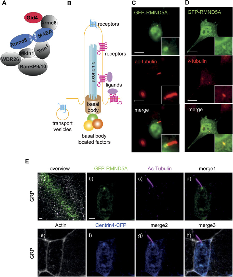
Fig. 1.
GID subunits colocalize to the basal body of mono-ciliated cells. (A) Schematic model of the vertebrate GID complex and its known subunits; RING domain-containing subunits are highlighted in blue, the substrate-recruiting factor GID4 is highlighted in red. (B) Schematic model of the primary cilium, composed of a basal body, axoneme and ciliary membrane. A large number of transporters, structural proteins, membrane receptors and basal body-located factors function in the primary cilium. (C,D) RMND5A localizes to the basal body of the primary cilium in NIH-3T3 cells. Cells were transfected with plasmids encoding GFP–RMND5A for 24 h and then further serum-starved (high glucose DMEM, 0.5% FCS) for an additional 24 h to induce ciliogenesis. After fixation, cells were stained for (C) acetylated tubulin (ac-tubulin) or (D) γ-tubulin to visualize the axoneme or the basal body of the primary cilium, respectively. Images were merged to identify overlapping signals (merge, yellow). Inset images show the magnification of a primary cilium and the basal body. Images shown in C and D are representative of seven and four images, respectively. Scale bars: 10 μm. (E) Rmnd5a localizes to basal bodies of motile mono-cilia of the GRP in X. laevis. mRNAs encoding GFP–Rmnd5a (green) and Centrin4–CFP (blue, centrioles/basal bodies) were injected at the four-cell stage. Embryos were fixed and stained at NF stage 17 to visualize cilia (magenta, ac-tubulin) and actin (white). Merge 1 shows a merged image of Eb and Ec; merge 2 shows a merged image of Ec and Ef; and merge 3 shows a merged image of Ee, Ef and Ec. Images are z-projections of confocal micrographs and represent five biological samples derived from one experiment. Scale bars: 20 μm (Ea), 3 μm (Eb–h).

