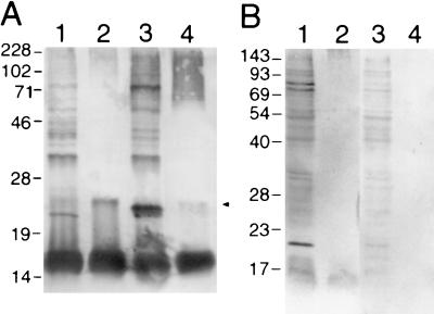
FIG. 4.
Western blot analysis of F. psychrophilum 259–93. (A) F. psychrophilum was grown in MAOB (lanes 1 and 2) or TYES (lanes 3 and 4). In lanes 1 and 3 are whole-cell lysates, and in lanes 2 and 4 are proteinase K digests of intact cells. The samples were separated by SDS–12% PAGE and reacted with rabbit anti-F. psychrophilum serum. Whole cells did not react with rabbit preimmune sera. (B) F. psychrophilum was grown in TYES and reacted with pooled rainbow trout convalescent anti-F. psychrophilum sera (lanes 1 and 2) and naïve rainbow trout sera (lanes 3 and 4). In lanes 1 and 3 are whole-cell lysates, and in lanes 2 and 4 are proteinase K digests of intact cells. Molecular mass markers (kDa) are indicated on the left of each blot. The arrow indicates a proteinase K-resistant antigen.

