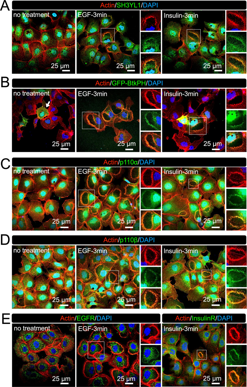
Fig. 6.
Receptor-mediated PI3K pathway at CDRs in Hep3B cells. A Representative confocal images of actin-SH3YL1 showing SH3YL1 is localized at CDRs (enlarge images). (B Representative confocal images of actin and GFP-BtkPH. GFP-BtkPH was expressed in Hep3B cells as a probe protein to identify PIP3. Confocal images showed recruitment of GFP-BtkPH to CDRs, indicating that PIP3 was generated at the structures. C and D Representative confocal images of actin-p110α (C) and actin p110β (D) showing p110 isoforms are localized at CDRs (enlarge images). E Representative confocal images of EGF receptor and insulin receptor. Both receptors are localized at CDRs in Hep3B (enlarge images)

