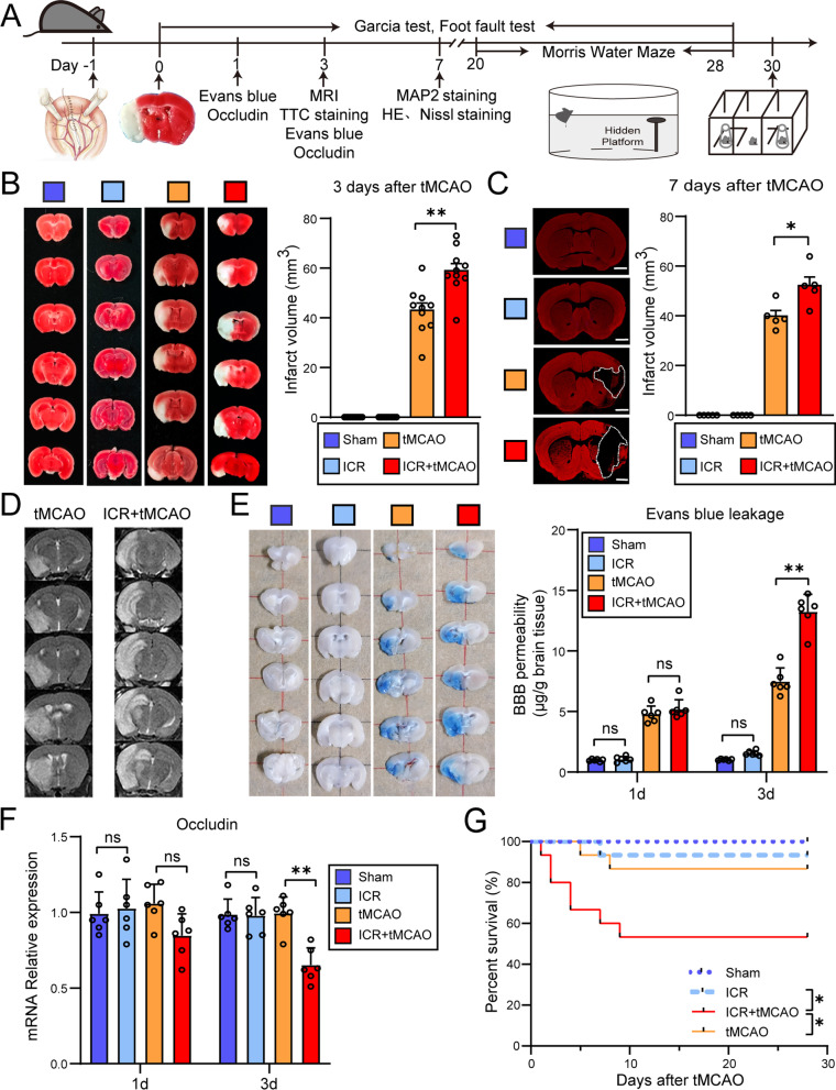
Fig. 1.
The perioperative stroke mice exhibit more severe cerebral ischemic injury than the stroke-only mice. A Flowchart illustrates the experimental design. B Representative images and quantification of TTC-stained coronal sections showing the infarct volume 3 days after stroke (n = 10/group). C Representative images and quantification of MAP2-stained coronal slices showing the infarct volume 7 days after stroke (n = 5/group). Scale bars = 1 mm. D Representative T2-weighted images (T2WI) of mice brain showing ischemic lesion. E Representative images and quantification of Evans blue extravasation (1 and 3 days after stroke) (n = 6/group). F Quantification of mRNA expression of tight junction protein occludin (1 and 3 days after stroke) (n = 6/group). G Kaplan–Meier estimates of survival until 28 days after tMCAO (n = 15/group). The sham mice were defined as the mice that underwent laparotomy without ICR. *P < 0.05, **P < 0.01, ns indicates nonsignificant

