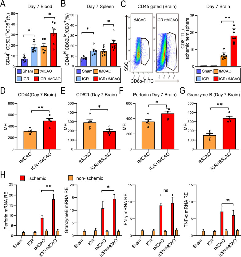
Fig. 3.
The activation and brain invasion of CD8+ T lymphocytes exacerbate ischemic brain injury in perioperative stroke mice. A, B Flow cytometry analysis on CD44hiCD62Llo percentage on CD8+ T lymphocytes in blood (A) and spleen (B) 7 days after stroke (n = 6–7/group). C Representative dot plots and an absolute number of brain-invading CD8+ T cells 7 days after stroke (n = 7/group). D–G CD44 (D), CD62L (E), Perforin (F), and Granzyme B (G) mean fluorescence intensity (MFI, arbitrary unit) of brain infiltrating CD8+ T cells 7 days after stroke (n = 5/group). H mRNA levels of cytokines Perforin, Granzyme B, IFN-γ, and TNF-α as measured by RT-PCR in ischemic and non-ischemic hemispheres (n = 5/group). The sham mice were defined as the mice that underwent laparotomy without ICR. *P < 0.05, **P < 0.01, ns indicates nonsignificant

