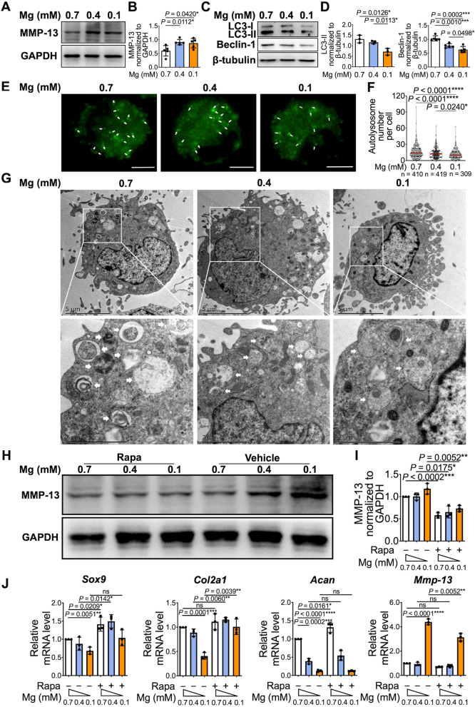
Fig. 2.
Reduced autophagy contributed to pro-catabolic and anti-anabolic effects after the exposure to magnesium deficient conditions. A Mouse chondrocytes were cultured with TNF-α under different magnesium conditions (0.7 mM, 0.4 mM, and 0.1 mM). Protein levels of MMP13 was detected by western blot. B The quantification of protein expression of MMP13 was done by densitometry analysis of the protein bands. Values were normalized to GAPDH (n = 5). C Protein level of LC3-II and Beclin-1 under different magnesium conditions were analyzed by western blot. D The quantification of protein expression of LC3-II and Beclin-1 was done by densitometry analysis of the protein bands. Values were normalized to β-tubulin (n = 5). E Chondrocyte imaging with DALGreen staining under different magnesium conditions. Autolysosomes were marked with white arrows. Scale bar: 5 μm. F Number of autolysosomes per cell was quantitated. G TEM analysis of autophagosomes in chondrocytes under different magnesium conditions. Autophagosomes were marked with white arrows. Scale bar: 5 μm. H Mouse chondrocytes were cultured with TNF-α under different magnesium conditions with or without rapamycin (Rapa) pretreatment. Protein levels of MMP13 was detected by western blot. I The quantification of protein expression of MMP13 was done by densitometry analysis of the protein bands. Values were normalized to GAPDH (n = 3). (J) Sox9, Col2a1, Acan, and Mmp-13 genes expression were assessed by qRT-PCR (n = 3). β-actin was used as housekeeping gene. Data were expressed as the mean ± SD and analyzed by one-way ANOVA test. (*P< 0.05, **P< 0.01, ***P< 0.001, ****P< 0.0001)

