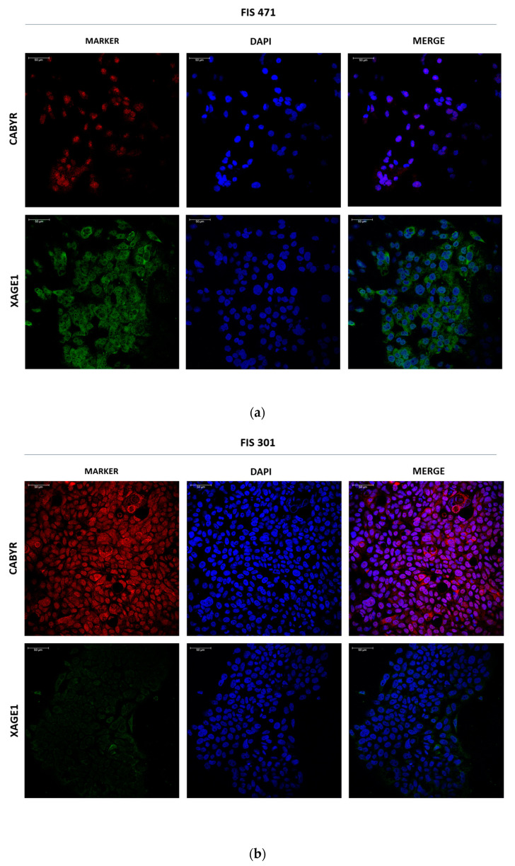
Figure 6.
Immunofluorescent staining of CABYR and XAGE1 in primary cultures. (a) Representative images of CABYR (red) and XAGE1B (green) in adherent-cultured cells from FIS 471 (LUAD). (b) Representative images of CABYR (red) and XAGE1B (green) in adherent-cultured cells from FIS 301 (LUSC). Cell nuclei are stained with DAPI (blue). Scale bar represents 50 μm.

