Figure 1.
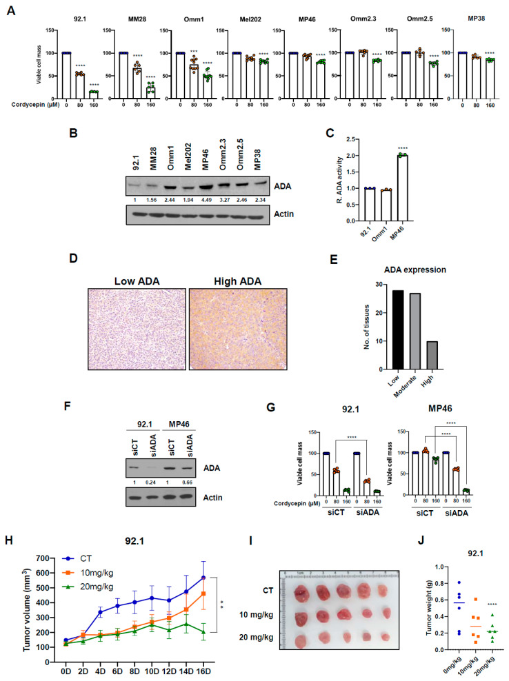
ADA decreases anticancer effects of cordycepin in uveal melanoma. (A) Growth of viable cell mass after cordycepin treatment. Uveal melanoma cells were treated with vehicle, 80, and 160 µM of cordycepin, and after 5 days incubation viable cell mass was measured using fluorescence. (B) Adenosine deaminase (ADA) protein expression levels varied uveal melanoma cells lines on western blots. (C) ADA enzymatic activity in uveal melanoma. (D,E) Immunohistochemical ADA protein expression in human uveal melanomas on tissue microarrays. (F,G), ADA protein expression (F) and cell growth (G) in 92.1 and MP46 uveal melanoma cells after siRNA-based knockdown. Values represent mean ± SD of experiments conducted in sextuplicate. **** p < 0.0001 by one-way ANOVA analysis of variance compared with control group. (H) Antitumor effects of cordycepin in mice bearing xenograft tumors of 92.1 uveal melanoma cells. (I) Pictures of xenografts from 92.1 uveal melanoma cells. (J) Tumor weight in vehicle, 10 mg/kg and 20 mg/kg cordycepin treatment group. Values represent mean ± SEM of experiments. CT, control; 10 mg/kg, cordycepin 10 mg/kg b.w. treatment; 20 mg/kg, cordycepin 20 mg/kg b.w. treatment. ** p < 0.01, *** p < 0.001, **** p < 0.0001 by one-way ANOVA analysis of variance compared with control group.

