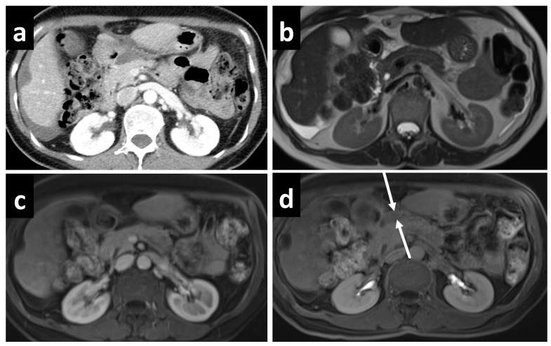
Figure 3.
A 61-year-old female with mucinous appendiceal neoplasm, a small amount of fluid is noted within the lesser sac on (a) CT and (b) T2W MRI. Enhancement within the fluid, however, is barely appreciated on the (c) portal venous phase of the MRI, but becomes clearly apparent on the (d) delayed MRI.

